THUNDER Imager 3D Cell Culture
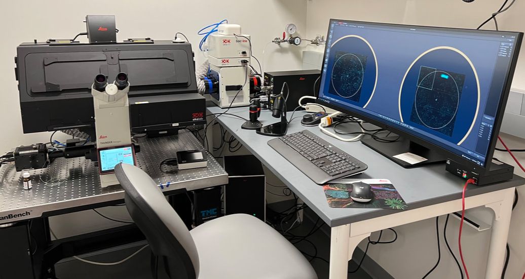
THUNDER Imager is a wide-field microscope that allows high-speed and high-quality 3D exploration of whole organisms, tissues, or cells. It features "Computational Clearing," which eliminates out-of-focus blur and enables shading correction. The microscope offers an environmental enclosure (temperature, humidity, and CO2 control), as well as an auto-focus to maintain samples at the focal plane over time. In addition, the instruments' “Spiral” option offers efficient tile scan imaging.
Applications:
390 nm, 440 nm, 475 nm, 510 nm, 555 nm, 575 nm, 635 nm, 747 nm.
Detectors: two QUAD cubes
Location: Longwood Center Basement Rm 1055D
Contact person: Paul Speliakos
Applications:
- Widefield imaging up to 8 channels.
- DIC
- Phase Contrast
- Polarizing microscopy
- Live-cell imaging
- Leica N Plan 5x/0.12 PH 0
- HC PL FLUOTAR 10x/0.32
- HC PL APO 20x/0.80
- HCX PL APO 40x/1.10 W CORR
- HC PL APO 63x/1.40-0.60 OIL
390 nm, 440 nm, 475 nm, 510 nm, 555 nm, 575 nm, 635 nm, 747 nm.
Detectors: two QUAD cubes
- DFT5 for DAPI/FITC/TRITC/Cy5
- CYR7 for DFP/YFP/Texas Red/Cy7
- Universal holding frame K
- 4-Slide insert
- Holding frame KM for Well Plates
Location: Longwood Center Basement Rm 1055D
Contact person: Paul Speliakos
Zeiss 980 Confocal/Spectral/Airyscan
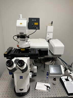
The LSM 980 microscope is a fully automated multimodal microscope that can be used for confocal, and spectral imaging. It is equipped with a unique AI Sampler Finder System. The Airyscan detector enables super-resolution and high-speed imaging. The LSM 980 is also equipped with Near-IR detectors that expand imaging spectra from UV to NIR.
Applications:
405 nm, 445 nm, 514nm, 561 nm, 594 nm, 639 nm
Detectors:
Location: Longwood Center Basement Rm 1055E
Contact person: Dr. Kun Huang
Applications:
- Confocal imaging
- Spectral imaging with linear unmixing
- Super-resolution imaging with resolution up to 90 nm using Airyscan
- Forster Resonance Energy Transfer (FRET)
- Fluorescence Recovery After Photobleaching (FRAP)
- Tile scan
- 5x air
- 10x air
- 20x air
- 25x multi-immersion (water, silicone oil, glycerin, oil) objective with 0.57 mm working distance
- 63x 1.4 oil
- Alpha Plan-APO 100x/1.46 Oil DIC VIS
405 nm, 445 nm, 514nm, 561 nm, 594 nm, 639 nm
Detectors:
- 34 Channel Quasar Detector Array
- Airyscan 2 Detectors
- Near-IR detectors (Filter set 8 deep red 640 +730 nm f/NIR and filer set 9 deep read 730 f/NIR)
- Scanning Stage (with Z-piezo)
Location: Longwood Center Basement Rm 1055E
Contact person: Dr. Kun Huang
Akoya PhenoCycler (CODEX) coupled with ZEISS Axio Observer 7
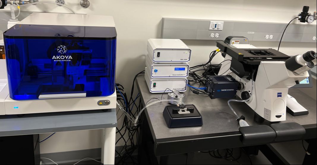
CODEX is a fast, unbiased, and highly-multiplexed imager for protein targets. It allows the phenotyping of millions of cells in complex tissue or environment as a single intact tissue section. Pushing the frontiers of spatial biology, our team offers services from tissue staining, to imaging, to machine-learning based cell segmentation and analysis.
Instrument:
- PhenoCycler (formerly CODEX) perfusion system
- ZEISS Axio Observer with Colibri 7 LED light Source
- 10x air
- 20x air
385 nm, 469 nm, 555 nm, 631 nm, 750 nm
Sample type: Mouse Fresh Frozen, Human FFPE
Operating software: Zen Software, Akoya Software
Location: Longwood Center Basement Rm 1055C
Contact person: Dr. Kun Huang
Image Analysis HP Workstation
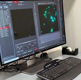
The HP workstation offers high-computing power for fast 3D reconstruction, image quantification and segmentation. Software installed:
- Zen desk (full version module)
- Fiji (ImageJ)
- CellProfiler (with CellPose and StarDist)
- CODEX processor
- QuPath
10x Genomics Xenium Analyzer
Xenium analyzer is a highly-multiplexed and sensitive imaging platform for RNA targets. It allows profiling transcriptome at single-cell and sub-cellular resolution.
Objectives:
- 20x water
Available Panels: 10x Genomics Panels with customized add-on options
Operating software: Xenium Ranger, Xenium Analyzer
Location: Longwood Center Basement Rm 1055C
Contact person: Dr. Kun Huang